
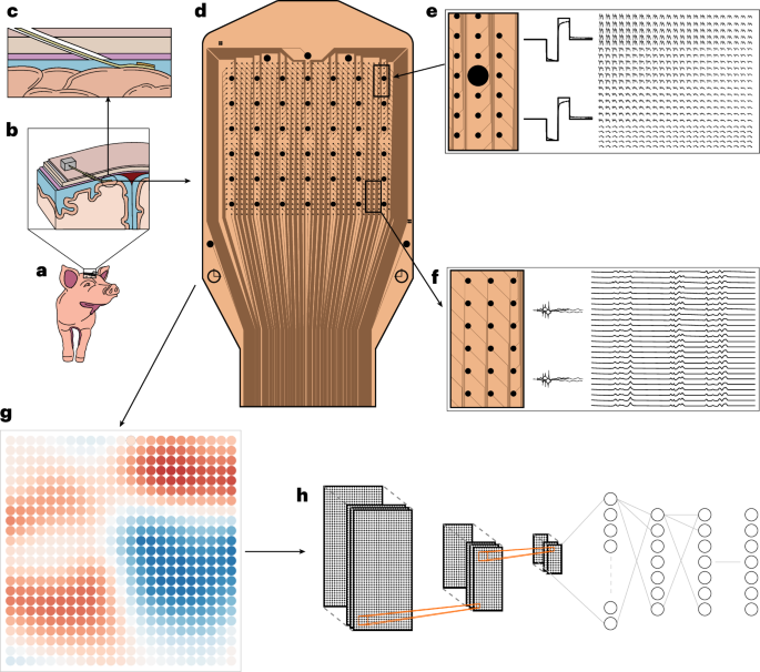
In 2025, an experimental study published in a Nature family journal demonstrated that a high‑density cortical microelectrode array containing 1,024 electrode channels can be delivered to the cerebral cortex through a very small incision in the skull and dura, enabling multimodal (recording + stimulation) function across a wide cortical area. The research team performed feasibility tests in pig models and in human neurosurgical patients, showing the system's trade‑offs between spatial coverage, signal modalities and surgical trauma.
The array's design emphasizes "scalability" and "minimal invasiveness". Rather than the conventional grid electrodes that require large craniotomies, the team introduced a flexible array through a small incision and deployed it over the cortical surface. The array contains thousands of microelectrodes and can simultaneously capture single‑unit spikes, local field potentials (LFPs) and lower‑frequency population activity. The described implantation path avoids the need for broad exposure of the cortical surface, significantly reducing surgical exposure time and tissue retraction.
In pig experiments, the authors deployed the array across large regions covering motor and sensory cortex and recorded signals with spatially fine‑grained differences. Electrodes served both as high‑resolution probes of neural activity and as stimulation sites to evoke or modulate local cortical activity. The paper notes the array supports bidirectional operation: recording for real‑time neural decoding and stimulation to trigger or correct neural circuits, which can be combined for closed‑loop interventions.
Attractive for clinical translation, the team also demonstrated an initial application in the operating room: short‑term implantation of the array in accessible regions during human neurosurgery for functional mapping and signal recording, illustrating the ability to acquire cross‑region neural activity and perform stimulation mapping within limited operative time. The authors emphasize this approach is not intended to replace all existing electrodes but provides a feasible surgical path for studies and clinical uses that require high‑channel data across larger cortical areas (for example, complex motor‑intention decoding, wide‑area epilepsy focus localization, or scalable brain–computer interfaces).
The paper details several engineering and clinical challenges, including long‑term tissue biocompatibility, stability of contact impedance over time, signal quality changes with implantation depth and coverage area, and the need for improvements toward fully percutaneous or catheter‑based delivery in clinical settings. The authors propose next steps including long‑term animal studies to validate electrode stability and stimulation safety windows and exploration of synergistic optimization with neural decoding algorithms.
This work appeals to early‑career researchers and developers because of its interdisciplinary approach: microelectronic array engineering, flexible materials, stereotactic minimally invasive surgical techniques and real‑time AI decoding algorithms. Together, these advances suggest future brain–computer interfaces could increase channel counts and functional complexity without a proportional rise in surgical burden. The study offers new methods and ideas for clinical translation of large‑scale, high‑resolution neural recording and intervention.